Unmasking pipefish otolith using synchrotron-based scanning X-ray fluorescence
Unmasking pipefish otolith using synchrotron-based scanning X-ray fluorescence
Haÿ, V., Berland, S., Medjoubi, K. et al. Unmasking pipefish otolith using synchrotron-based scanning X-ray fluorescence. Scientific Reports 13, 4794 (2023). https://doi.org/10.1038/s41598-023-31798-z
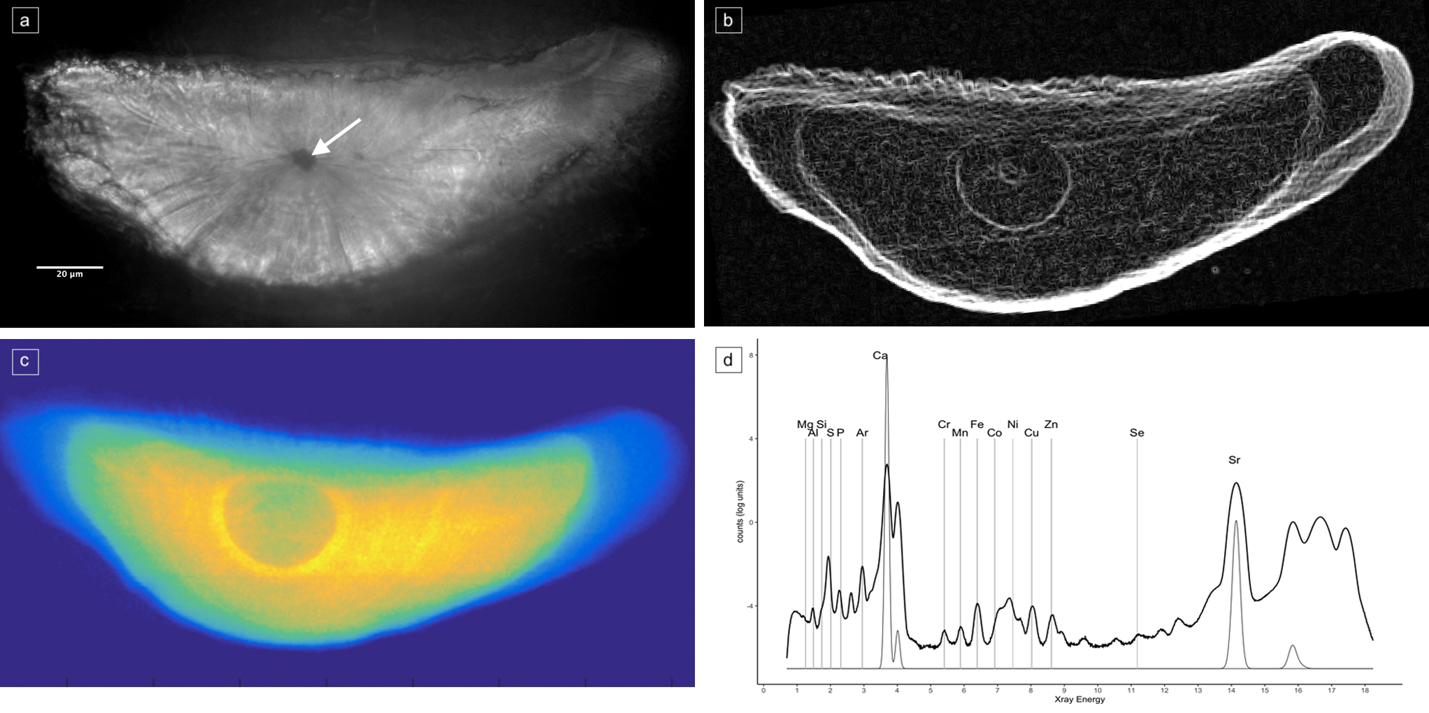
Scientists use otoliths to trace fish life history, especially fish migrations. Otoliths incorporate signatures of individual growth and environmental use. For many species, distinct increment patterns in the otolith are difficult to discern; thus, questions remain about crucial life history information. To unravel the history of such species, we use synchrotron-based scanning X-ray fluorescence. It allows the mapping of elements on the entire otolith at a high spatial resolution. It gives access to precise fish migration history by tagging landmark signature for environmental transition and it also characterises localised growth processes at a mineral level. Freshwater pipefish, which are of conservation concern, have otoliths that are small and fragile. Growth increments are impossible to identify and count; therefore, there is a major lack of knowledge about their life history. We confirm for the first time, by mapping strontium that the two tropical pipefish species studied are diadromous (transition freshwater/marine/freshwater). Mapping of other elements uncovered the existence of different migratory routes during the marine phase. Another major breakthrough is that we can chemically count growth increments solely based on sulphur signal as it is implicated in biomineralization processes. This novel method circumvents reader bias issues and enables age estimation even for otoliths with seemingly untraceable increments. The high spatial resolution elemental mapping methods push back limits of studies on life traits or stock characterisation.
BOREA contact: Vincent Haÿ, vincent.hay@etu.sorbonne-universite.fr
Picture title: Application of synchrotron X-ray fluorescence (XRF) on freshwater pipefish otoliths. (A) Polarized light microscopy of Microphis nicoleae otolith. (B) Delimitation of zonations in the otolith. (C) Elemental map of strontium. (D) Elemental composition identified in the otolith from XRF method. © V. Haÿ


