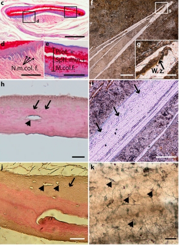
The presence of a pulmonary organ that is entirely covered by true bone tissue and fills most of the abdominal cavity is hitherto unique to fossil actinistians. Although small hard plates have been recently reported in the lung of the extant coelacanth Latimeria chalumnae, the homology between these hard structures in fossil and extant forms remained to be demonstrated. Here, we resolve this question by reporting the presence of a similar histological pattern–true cellular bone with star-shaped osteocytes, and a globular mineralisation with radiating arrangement–in the lung plates of two fossil coelacanths (Swenzia latimerae and Axelrodichthys araripensis) and the plates that surround the lung of the most extensively studied extant coelacanth species, L. chalumnae. The point-for-point structural similarity of the plates in extant and fossil coelacanths supports their probable homology and, consequently, that of the organ they surround. Thus, this evidence questions the previous interpretations of the fatty organ as a component of the pulmonary complex of Latimeria.
_____________________
*The homology and function of the lung plates in extant and fossil coelacanths. Cupello, C., Meunier, F.J., Herbin, M., Janvier, P., Clément, G. & Brito, P.M. (2017) The homology and function of the lung plates in extant and fossil coelacanths. Scientific Reports, 7 :9244, DOI :10.1038/s41598-017-09327-6
BOREA contact : François J. Meunier, francois.meunier@mnhn.fr
Picture title: Extant and fossil coelacanth lung plates. (c) Histological thin section of a Latimeria chalumnae lung plate (adult specimen CCC 5, azocarmin coloration). (d) Close-up of boxed area in (c), focussing on collagen fibres at the extremity of the plate, which corresponds to the non-mineralised region. (e) Close-up of boxed area in (c), focussing on mineralised portion of the plate comprising proteoglycans, collagenous fibres, and spheritic mineralisation. (f) Ground cross-thin section of Axelrodichthys araripensis (UERJ-PMB 143) lung plate showing central artefactual fracture. (g) Close-up of boxed area in (f), focussing on weakness zone. (h) Histological thin section of a Latimeria chalumnae lung plate (haematoxylin/eosin coloration, adult specimen CCC 24); black arrows point to mineralised globules with a radiating arrangement; arrowhead points to osteocyte lacunae. (i) Ground cross-thin section of Axelrodichthys araripensis lung plate (UERJ-PMB 143); black arrows point to mineralised globules with a radiating arrangement. (j) Histological thin section of an ossified plate of the extant coelacanth Latimeria chalumnae (adult specimen CCC 24); arrowheads point to star-shaped osteocytes and arrow to canaliculi. (k) Ground cross-thin section of an Axelrodichthys araripensis lung plate (UERJ-PMB 143); arrowheads point to star-shaped osteocytes with ramified canaliculi for cytoplasmic processes. Ext. surf., external surface; Int. surf., internal surface; Min., mineralised portion; m. col. f., mineralised collagen fibres; n. m. col. f., non-mineralised collagen fibres; n. min., non-mineralised portion; prot, proteoglycans; sph. min., spheritic mineralisation; w. z., weakness zone. Scale bars, 500 µm (a–c,f) 100 µm (d,e,i) 2 µm (g) 20 µm (h) 50 µm (j,k).
